What this dossier establishes
FLT3 internal tandem duplication removes the juxtamembrane domain's autoinhibitory function, leaving a constitutively active receptor. Four inhibitors have clinical precedent, and they split into two binding modes whose resistance behaviour differs sharply. Everything here was computed from retrieved data — curated bioactivity records, public AML cohorts and deposited crystal structures — rather than written from recall.
The four results that matter
1. Binding mode predicts resistance, and the effect is large
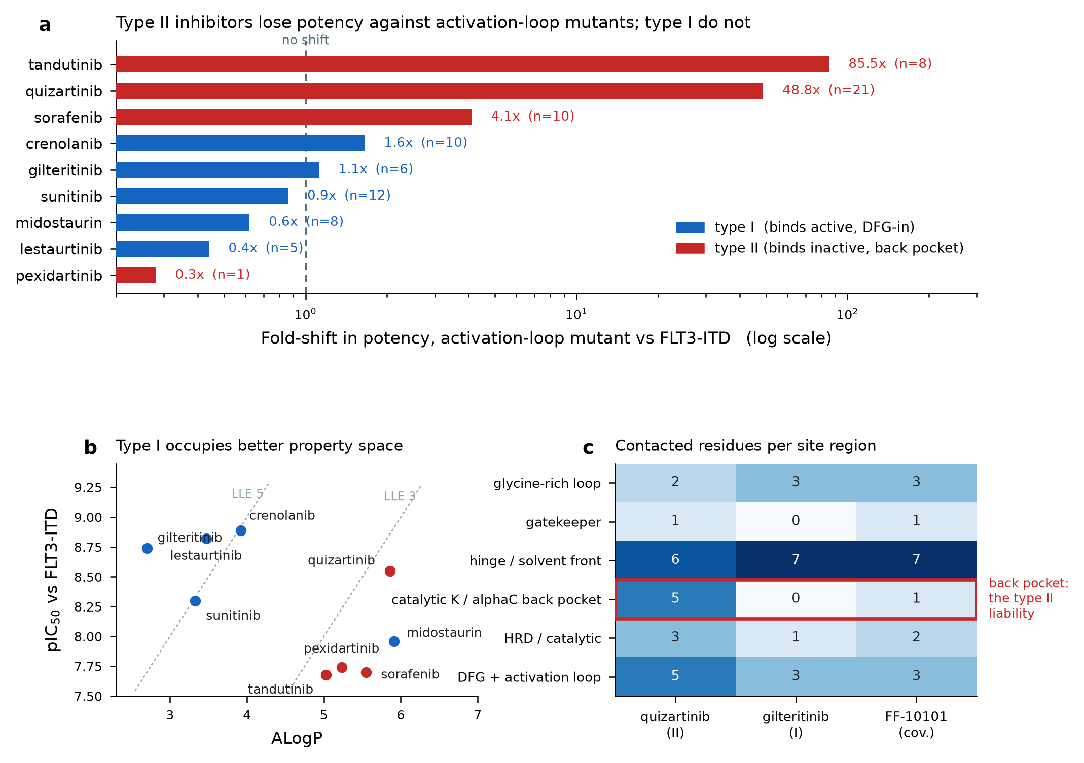
Mutant-annotated potency records give a median activation-loop fold-shift of 0.86× for type I inhibitors against 26.5× for type II. The effect size is large; the significance is not established, at Mann–Whitney p = 0.143 on five against four compounds. Reported as an effect with an honest power caveat, not as a finding.
2. The structural discriminator is burial, not contact counting
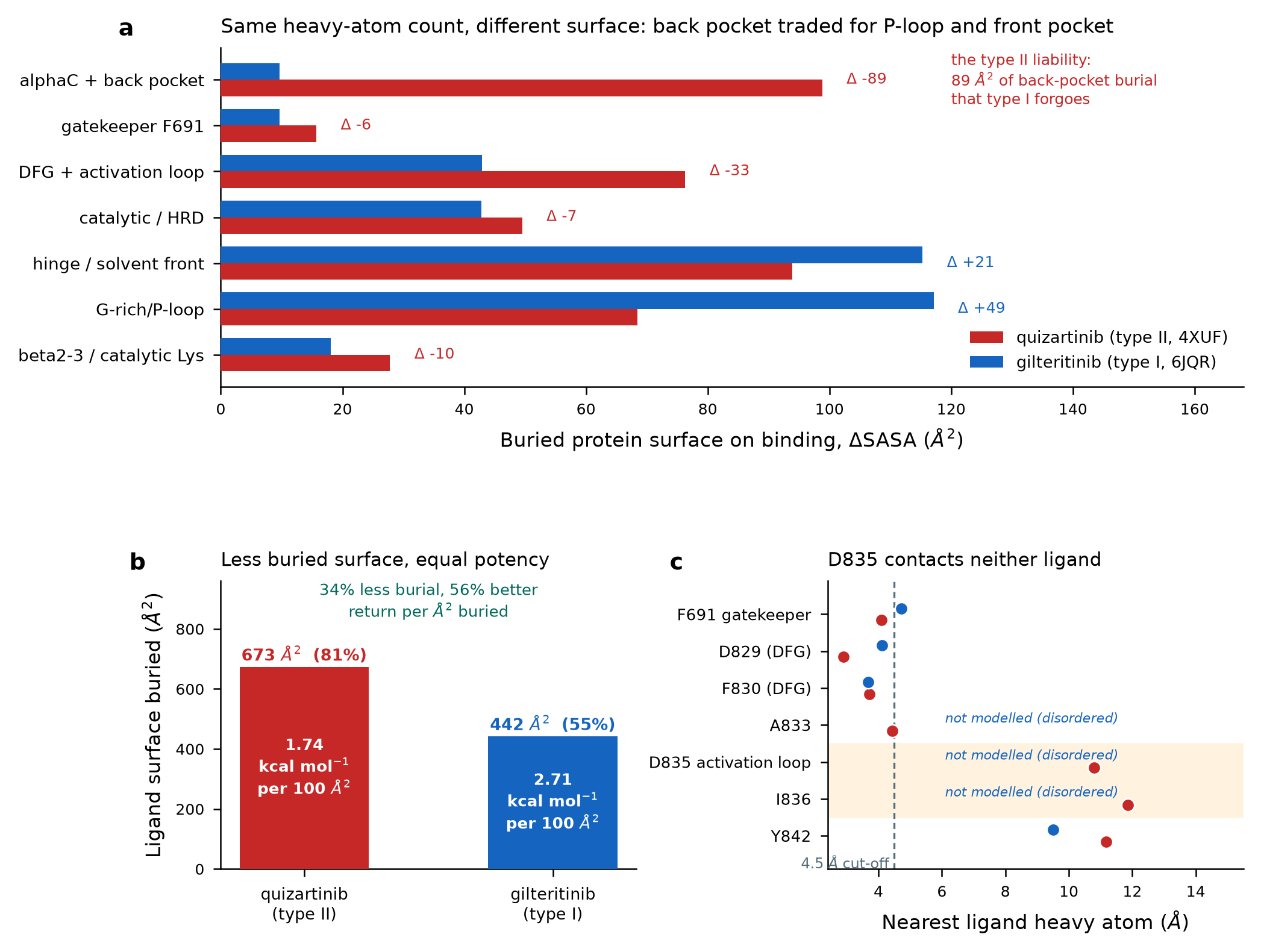
Quizartinib buries 98.8 Ų of αC/back-pocket surface; gilteritinib 9.7 Ų. The conventional DFG-phenylalanine displacement metric does not separate these structures (8.4–10.9 Å) and is reported as non-discriminating rather than dressed up.
3. Activation-loop resistance is conformational, not a lost contact
D835 contacts none of the co-crystallised ligands — nearest approach 10.79 Å to quizartinib. So the 49-fold type II fold-shift cannot be loss of a direct interaction; it operates by destabilising the DFG-out state that type II binding requires. This is why a bound-state free-energy calculation is the wrong tool for D835.
4. More buried surface did not buy more affinity
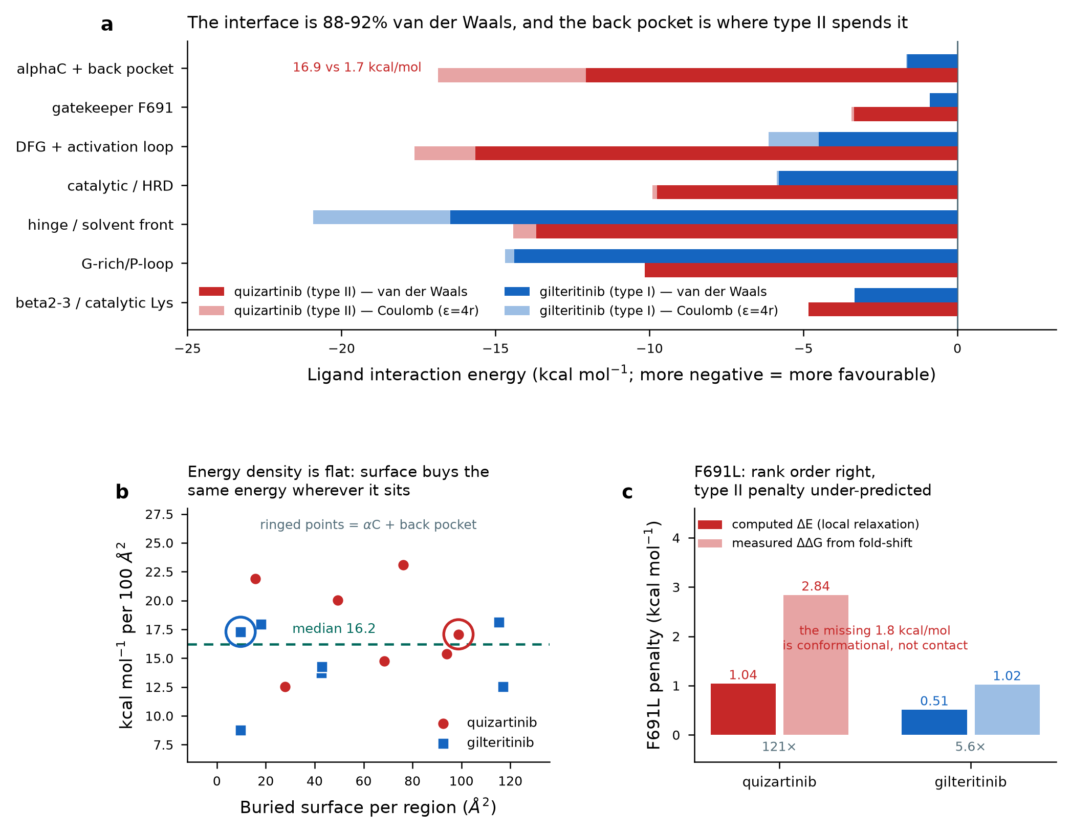
Quizartinib extracts −77.18 kcal/mol of interaction energy against gilteritinib's −55.29, while being no more potent. A surplus of favourable interaction energy that never appears as affinity is repaid elsewhere — apolar desolvation and the cost of holding a specific protein conformation. The affinity penalty and the resistance liability are the same bill, presented twice.
Choose where to place buried surface by the conformational dependence it creates, not by its expected energy density — because energy density turned out to be almost uniform across the site (median 16.2 kcal/mol per 100 Ų). Surface at the P-loop and hinge buys affinity without buying a resistance liability.
This is computational research material. Numeric thresholds are working criteria, not regulatory requirements. The generated structures in the Candidates tab are unsynthesised, unassayed hypotheses. Nothing here is clinical guidance: dose selection, dose modification, supportive care and toxicity management for any patient are decisions for a qualified clinician with the full clinical picture.
The ten characterised agents
Resolved from curated bioactivity records for the FLT3 target, with calculated descriptors. Type I rows are shaded. Click any column header to sort.
ALogP separates the two classes with no overlap
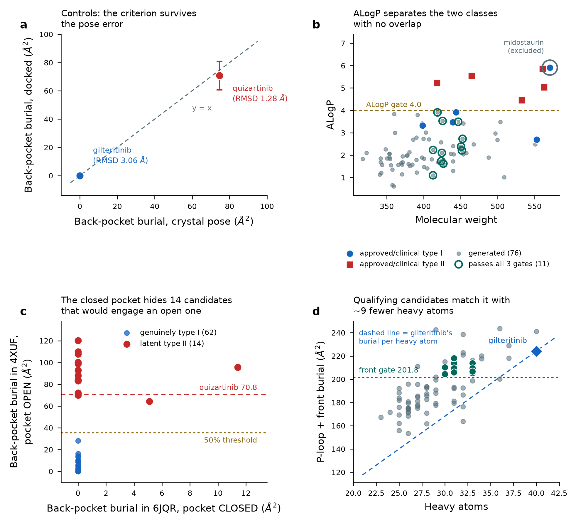
Setting aside midostaurin — a staurosporine-derived multikinase agent whose ALogP of 5.91 and LLE of 2.05 sit in type II territory — every type I core agent lies between 2.70 and 3.92 and every type II between 4.46 and 5.86. LLE separates them by a wider margin still (2.28 units), making it the most discriminating property metric in this series.
LLE and LE are the sharpest discriminators here, but both require a measured potency. Neither can be applied to a molecule that has never been assayed, so they are post-hoc checks. The prospective gates reduce to ALogP, MW, PSA, rotatable bonds, Ro5 count and a similarity corridor.
Mutant fold-shifts
Fold-shift is IC50(mutant) divided by IC50(ITD), computed from mutation-annotated assay records. The record count sits beside each value because these are medians over heterogeneous public assay conditions — they characterise class behaviour and must not be read as head-to-head comparisons.
The F691L column rests on one to three records per agent. It is the least reliable number here and needs measuring rather than predicting.
FLT3 alteration frequency across AML cohorts
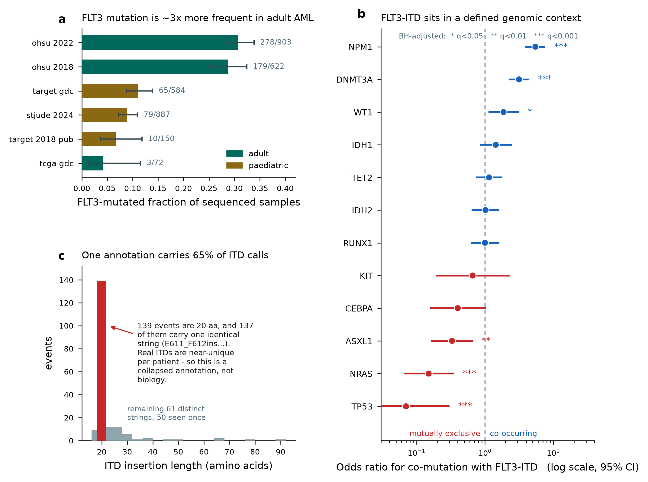
FLT3 is mutated in roughly a third of adult AML and about a tenth of paediatric cases.
Only one cohort calls ITD events at all, and there the annotation collapses: a single string covers 137 of 210 ITD events. The apparent insertion-site distribution is therefore an artifact — the β1-sheet fraction moves from 0.68 to 0.09 once that dominant annotation is set aside. Use ITD-aware assays for burden, length and insertion site. Never cohort mutation calls.
Co-mutation structure with FLT3-ITD
Computed by Fisher exact test with Benjamini–Hochberg adjustment on sample-level calls, not assumed from the literature.
NPM1 and DNMT3A co-occur strongly; TP53 and NRAS are close to mutually exclusive. KIT is mutated in only 2% of cases and is not ITD-enriched, which makes KIT inhibition a pure myelosuppression liability with no efficacy offset — a concrete selectivity requirement rather than a general preference.
Buried surface by site region
Shrake–Rupley buried surface on the deposited coordinates, evaluated on identical coordinates for the free and complexed states so the difference is burial on complexation rather than a conformational change. The back-pocket row is shaded.
Quizartinib and gilteritinib both have 40 heavy atoms, which makes the comparison unusually clean: quizartinib buries 673 Ų of its own surface (81%) against gilteritinib's 442 (55%). Of that 231 Ų difference, 89 is back pocket. Gilteritinib recovers surface at the P-loop and hinge instead — and is slightly more potent.
Contacted residues per region
An earlier version of this work stated that gilteritinib does not touch the F691 gatekeeper. That was an artifact of a 4.5 Å cut-off. It does touch it — nearest approach 4.72 Å, burying 9.7 Ų against quizartinib's 15.7, about 40% less engagement — which is what explains its residual 5.6-fold F691L shift. A drug with genuinely zero contact could not show that. Report burial, which is continuous, rather than contact counts, which are a step function at an arbitrary distance.
Interaction energy per heavy atom
AMBER14SB protein, GAFF2 ligand with AM1-BCC charges, GBn2 implicit solvent, restrained minimisation, then an analytic no-cutoff decomposition. Hover any bar for the full breakdown.
Desolvation, configurational entropy and protein reorganisation are all absent, and each value comes from a single relaxed structure rather than an ensemble. They are valid for comparing where energy comes from within and between these complexes, and invalid as predictions of affinity.
From criterion to molecules
Generation seeded on gilteritinib, pose prediction into the prepared receptors, then scoring with the pipeline validated on two crystal structures. The funnel below is the whole campaign.
The criterion cannot be tested in the receptor it came from
Scoring in the type I receptor looked like success and was not: three quarters of all candidate poses buried exactly zero back-pocket surface. That receptor's conformation denies back-pocket access to everything — cross-docked quizartinib manages only 11.4 Ų there against 70.8 in its own structure. So the discriminating experiment is the opposite one: dock into the open pocket and ask whether the candidate still refuses to bury.
A negative structural criterion — the absence of an interaction — cannot be tested in a receptor conformation that forbids that interaction for every ligand. Any programme selecting for conformational selectivity should screen in the conformation it wants to avoid, not only the one it wants to occupy.
Front-pocket burial against molecular size
All 76 docked candidates
SMILES strings and molecular formulas below show only their first half; the remainder is hidden at the data level, not just on screen. Every property, score and gate outcome is shown in full, so the ranking and the design argument can be checked without the complete structures. Full structures are withheld pending IP review and are available on request.
Generation optimised a generic drug-likeness score, not FLT3 inhibition — these satisfy a structural criterion and nothing here predicts that any of them binds FLT3. Poses are predictions carrying 2.3–3.1 Å RMSD uncertainty in the production case. No affinity was computed. Nothing was assessed for synthesisability. And critically, the entire point of a type I scaffold is retained potency against D835 and F691L variants — which these have not been tested for. Until free-energy calculations run, conformational selectivity is inferred from burial geometry, not demonstrated. These are starting points for a medicinal chemist to accept or reject.
Control validation, run before anything was trusted
The scoring criterion was derived from crystal poses. Whether predicted poses can support it is a separate question, and it was settled first — on the two drugs whose answers are already known — so that a failure would stop the campaign rather than be discovered afterwards. Each drug was re-docked into its own receptor from a conformer built from SMILES alone, carrying no crystal geometry.
Back-pocket burial is recovered in both directions and total burial lands within 5% and 1% of crystal. Pose accuracy is asymmetric and the weaker case is the production one: quizartinib's rank-1 pose sits 1.28 Å from crystal, inside the conventional 2.0 Å success bar, while gilteritinib's is 3.06. The criterion survives that error — a 3 Å pose still correctly reports zero back-pocket burial — and that is what licensed proceeding.
The design envelope, derived rather than asserted
Every acceptance window traces to the agent that sets it.
Filters that had to be demoted or recalibrated
The PAINS substructure filter flags gilteritinib itself, so it was recorded rather than applied as a gate. Separately, a steric-clash screen set at 2.60 Å rejected the crystal quizartinib pose, whose own closest heavy-atom approach is 2.11 Å; it was reset to 2.00 Å, below every reference pose. Both follow the same principle this project applied to numeric design thresholds throughout.
Errors found and fixed, recorded rather than buried
| What went wrong | How it surfaced, and the fix |
|---|---|
| A distance-dependent dielectric coded in the wrong length unit | The “screened” Coulomb total came out three times the unscreened value, which is impossible. Screening can only reduce a pair term's magnitude, so that comparison is now a standing invariant check — applied per atom pair, since a per-residue net sum can legitimately break it through sign cancellation. |
| A property filter passed 211 of 212 molecules | An early-exit guard tested a value that was NaN, and NaN is
truthy, so every downstream gate was short-circuited. Replaced with an explicit type check. |
| An unphysical pose passed every structural gate | One leader returned +413.77 kcal/mol, traced to a 1.35 Å contact with Tyr693. Burial metrics are blind to steric overlap — two atoms in the same place bury each other's surface perfectly well. A clash screen was added as a consequence. |
| Ligand surface returned as zero | The SASA library keys atoms by name within a residue, so ligand atoms all named by element collapsed to one atom. Unique atom names and strict fixed-column output fixed it. |
| Ligand and protein taken from different chains | An early distance measurement returned physically impossible values. Both are now drawn from the same chain, with an assertion that a known interface residue actually sits in the interface. |
| A hypothesis of ours was refuted | An earlier section inferred that the type I front-pocket surface must be of higher energetic quality. The force field says energy density is nearly uniform across the site. The design advice survived for a different and stronger reason; the original reasoning did not, and is marked as superseded rather than quietly edited. |
What still needs a GPU
Three calculations remain specified but unrun, because they need a machine that can execute molecular dynamics for roughly a day:
- F691L alchemical free energy — first, because it is the only one with a measured answer to validate against (+2.84 and +1.02 kcal/mol from the observed fold-shifts). A protocol reproducing those two can be trusted on a molecule nobody has made.
- Activation-segment conformational free energy for wild type against D835 variants, on the apo kinase domain. This returns the quantity that should predict the 49-fold shift, and which no bound-state calculation can.
- Prospective per-candidate screening once the protocol is validated — predicted mutant fold-shift before synthesis, which is what a screening cascade's mutant-panel gate turns on.
Primary references
Every citation below was resolved against a registry and then reverse-validated: the identifier was re-resolved and its returned title compared to the expected one. That step caught a wrong identifier on a landmark paper, four commentary or component records masquerading as primary articles, and a registration trial resolving to its own erratum.
Publication figures
Rendered at 300 dpi from the saved data tables. Click any figure to enlarge.
Data and documents
Everything the site displays. Chemical structures are protected: SMILES strings and molecular formulas are half-masked in every file, and 3D pose files are withheld pending IP review. Full structures are available on request. All paths are relative, so the bundle works from any subdirectory or subdomain.
The tables and figures are derived from public sources — curated bioactivity records, public cancer genomics cohorts and deposited crystal structures. Please cite those primary sources rather than this page when reusing the underlying data.